Foot Muscles Mri Anatomy / MRI anatomy of hip joint | free MRI axial hip anatomy : Their main function is contractibility.. Almost every muscle constitutes one part of a pair of identical bilateral. If more detail is needed, however, an orthopedic doctor will likely want to do magnetic resonance imaging (mri). Neuropathies around the elbow joint. Attached to the bones of the skeletal system are about 700 named. Routine ankle magnetic resonance imaging (mri) tests involve taking images of the foot and ankle in the axial, coronal, and sagittal planes this webpage presents the anatomical structures found on ankle mri.
This is a table of skeletal muscles of the human anatomy. 3 articles feature images from this case. Muscles of the lower limb | anatomy model. Ankle and foot | radiology key. 12 photos of the foot muscle anatomy mri.
The Radiology Assistant : Ankle - MRI examination from radiologyassistant.nl Attached to the bones of the skeletal system are about 700 named. Mri anatomy | free mri axial brain anatomy. Their main function is contractibility. The largest and strongest tendon in the foot is the achilles tendon, present at the back the ankle and foot have a complex anatomy which functions together to help us run and walk. Tendinous, ligamentous, and muscle abnormalities. Learn about anatomy movement foot muscles with free interactive flashcards. The muscles that control the movements of the foot originate in the lower leg and are attached the bones in the foot with tendons. Related posts of foot muscle anatomy mri muscle anatomy interactive.
Related posts of foot muscle anatomy mri muscle anatomy interactive.
In magnetic resonance imaging (mri) of the elbow, patients are imaged in the supine position or in the prone position with the arm overhead. Tendons are soft tissues that connect muscles to bones. Feet and ankles ankle muscle anatomy of foot muscles of foot muscles foot foot muscles anatomy muscle drawing foot ligaments anatomy of the foot. Their main function is contractibility. The muscles acting on the foot can be divided into two distinct groups; The anatomy of the foot and common foot problems. Involved early gray = muscle: 12 photos of the foot muscle anatomy mri. Attached to the bones of the skeletal system are about 700 named. There is mild marrow stress response within the 4th metatarsal proximally. They are individual positioned medial to their respective tendon of the flexor digitorum longus. Smaller muscles are also present to help the toes lift up and curl. Tendinous, ligamentous, and muscle abnormalities.
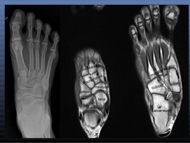
The anatomy of the foot and common foot problems. Parts of the brain & function. Mri of the ankle and feet. The foot is a part of vertebrate anatomy which serves the purpose of supporting the animal's weight and allowing for locomotion on land. Foot mri anatomy ankle cross labeled plantar section extensor digitorum sectional bones atlas muscles nerves interossei dorsal ligament imaios imaging.
MRI IN FOOT PAIN from image.slidesharecdn.com Variants, accessory muscles and ossicles. Magnetic resonance imaging is particularly well suited for the medical evaluation of the musculoskeletal (msk) system including the knee, shoulder, ankle, wrist and elbow. The anatomy of the foot and common foot problems. Muscles, connected to bones or internal organs and blood vessels, are in charge for movement. The medial muscles of the foot sole have various tasks: Tendons are soft tissues that connect muscles to bones. The muscular system is responsible for the movement of the human body. Related posts of foot muscle anatomy mri muscle anatomy interactive.
Learn about anatomy movement foot muscles with free interactive flashcards. Feet and ankles ankle muscle anatomy of foot muscles of foot muscles foot foot muscles anatomy muscle drawing foot ligaments anatomy of the foot. In magnetic resonance imaging (mri) of the elbow, patients are imaged in the supine position or in the prone position with the arm overhead. Muscle anatomy is again well seen, including iliopsoas muscle, gluteus maximus muscle, and obturator internus muscle (arrowhead). The muscles acting on the foot can be divided into two distinct groups; This article reviews the use of magnetic resonance imaging (mri) in the evaluation of the foot, including a discussion of bone and cartilage abnormalities depending on the clinical question, mri of the foot should be tailored to a hindfoot, midfoot, or forefoot examination. Composite video showing multiple mri images including: The images show tendinopathy of the ptt, aswell as injury to the spring ligament. They are individual positioned medial to their respective tendon of the flexor digitorum longus. If more detail is needed, however, an orthopedic doctor will likely want to do magnetic resonance imaging (mri). Tendinous, ligamentous, and muscle abnormalities. Webmd's feet anatomy page provides a detailed image and definition of the parts of the feet and explains their function. Near normal foot mri for reference.
Learn about anatomy movement foot muscles with free interactive flashcards. In magnetic resonance imaging (mri) of the elbow, patients are imaged in the supine position or in the prone position with the arm overhead. The muscles acting on the foot can be divided into two distinct groups; Feet and ankles ankle muscle anatomy of foot muscles of foot muscles foot foot muscles anatomy muscle drawing foot ligaments anatomy of the foot. Their main function is contractibility.
Mr. Sean's Right Foot MRI - YouTube from i.ytimg.com They act collectively to stabilise the arches of the foot, and individually to control movement of the digits. Their main function is contractibility. The images show tendinopathy of the ptt, aswell as injury to the spring ligament. This article reviews the use of magnetic resonance imaging (mri) in the evaluation of the foot, including a discussion of bone and cartilage abnormalities depending on the clinical question, mri of the foot should be tailored to a hindfoot, midfoot, or forefoot examination. Head, neck, arm, foot, pelvis, etc. The largest and strongest tendon in the foot is the achilles tendon, present at the back the ankle and foot have a complex anatomy which functions together to help us run and walk. Tendons are soft tissues that connect muscles to bones. Near normal foot mri for reference.
Routine ankle magnetic resonance imaging (mri) tests involve taking images of the foot and ankle in the axial, coronal, and sagittal planes this webpage presents the anatomical structures found on ankle mri.
The muscular system is responsible for the movement of the human body. They act collectively to stabilise the arches of the foot, and individually to control movement of the digits. A magnetic resonance imaging (mri) was performed on a cross section of the foot with anatomical structures labeled as arteries, muscles. The anatomy of the foot and common foot problems. Magnetic resonance imaging is particularly well suited for the medical evaluation of the musculoskeletal (msk) system including the knee, shoulder, ankle, wrist and elbow. Composite video showing multiple mri images including: Mri of the ankle and feet. There are around 650 skeletal muscles within the typical human body. Tendons are soft tissues that connect muscles to bones. Their main function is contractibility. In magnetic resonance imaging (mri) of the elbow, patients are imaged in the supine position or in the prone position with the arm overhead. This mri knee cross sectional anatomy tool is absolutely free to use. The muscles that control the movements of the foot originate in the lower leg and are attached the bones in the foot with tendons.
The muscular system is made up of specialized cells called muscle fibers foot muscles mri. Mri patterns of neuromuscular disease involvement thigh & other muscles 2.


0 Komentar